MWA in Fibroids

'Save your uterus'

MWA in Liver MWA in Thyroid MWA in Fibroids MWA in Kidney MWA in Lung

Home > Solutions - Microwave Ablation - MWA in Fibroids

Introduction of Microwave Ablation in Fibroids

Uterine fibroids are benign tumors that occur most commonly (70-80%) in women of childbearing age [1] . Fibroids impose a significant psychological burden on affected women. Patients with symptoms often must be treated to alleviate those symptoms [2].

Treatment options for fibroids include hysterectomy, myomectomy, hysteroscopic resection and minimally invasive approaches. Surgical treatment remains the reference standard in the treatment of uterine fibroids; however, it may cause severe trauma and undesirable comorbidity [3].

Actually, not every patient is suitable for the surgical treatment. And improvement in quality of life is a major concern for women evaluating options for fibroids treatment [4]. Thus, increasing attention has been paid to the study of minimally invasive or non-invasive therapies with uterine preservation such as Microwave Ablation [5].


How Microwave Ablation Works

Microwave ablation refers to the use of the electromagnetic methods for inducing tumor destruction by using devices with frequencies of at least 900 MHz to produce tissue-heating effects [4]. Currently, 2450MHz is the frequency used by mainstream products worldwide.

How Effective Microwave Ablation is?

In 2011, a report comes from the PLA General Hospital of China shows forty patients underwent the MWA treatment. The baseline fibroids volume was 14.6 to 341.1 cm3 (mean 140.1 ± 87.4cm3). The shrinkage rate of the fibroids was 61.8%, 78.7%, 73.2% and 93.1% at 3, 6, 9, and 12 months after ablation, respectively [10].

In 2016, Chinese doctors released a multi-center research report. It shows a total of 311 patients (405 leiomyomas) from eight treatment centers underwent the treatment (age, 29-55 years; mean ± SD, 41 ± 5.11 years). The mean ablation rate was 86.6% (54.0-100%). The mean reduction rate was 63.5%, 78.5% and 86.7% at 3-, 6- and 12-months posttreatment, respectively. The symptom severity score (SSS) and health-related quality of life scores were also significantly improved posttreatment compared with before treatment (p=.000) [11].

In 2022, Swedish doctors released a study report which involving 16 patients. Most patients (82%) reported improvement up to 3 years after treatment. Out of 42 treated fibroids, 35 (83%) continued to shrink over time with median relative volume reduction of 77% (IQR39-95) [12]. 

In conclusion, MWA is an effective minimally invasive treatment for symptomatic uterine fibroids that can significantly improve the quality of life of patients.

1698385469049

Clinical Cases

MR images of hypointense leiomyomas before and after percutaneous MWA (PMWA) in a 32-year-old female, measuring 5.5x5.2x 4.9 cm and 5.4x5.0×4.6 cm, respectively. (a) T2 weighted sagittal MR image of a patient before PMWA shows two hypointense leiomyomas (arrows). (b) Two hypointense leiomyomas show minor enhancement on contrast- enhanced MRI (ceMRI) before PMWA (arrows). (c) ceMRI of the same patient after PMWA; the length and height of the non-perfusion area of a leiomyoma were measured on sagittal ceMRI. (d) ceMRI of the same patient after PMWA; the width of the non-perfusion area of a leiomyoma was measured on axial ceMRI [13].


Advantages

Reference List

1. Baird, D. D., Dunson, D. B., Hill, M. C., Cousins, D., & Schectman, J. M. (2003). High cumulative incidence of uterine leiomyoma in black and white women: ultrasound evidence. American journal of obstetrics and gynecology, 188(1), 100-107.

2. Levy, B. S. (2008). Modern management of uterine fibroid. Acta obstetricia et gynecologica Scandinavica, 87(8), 812-823.

3. Harding, G., Coyne, K. S., Thompson, C. L., & Spies, J. B. (2008). The responsiveness of the uterine fibroid symptom and health-related quality of life questionnaire (UFS-QOL). Health and quality of life outcomes, 6, 1-8.

4. Go, V. A. A., Thomas, M. C., Singh, B., Prenatt, S., Sims, H., Blanck, J. F., & Segars, J. H. (2020). A systematic review of the psychosocial impact of fibroids before and after treatment. American journal of obstetrics and gynecology, 223(5), 674-708.

5. Kanaoka, Y., Yoshida, C., Fukuda, T., Kajitani, K., & Ishiko, O. (2009). Transcervical microwave myolysis for uterine myomas assisted by transvaginal ultrasonic guidance. Journal of Obstetrics and Gynaecology Research, 35(1), 145-151.

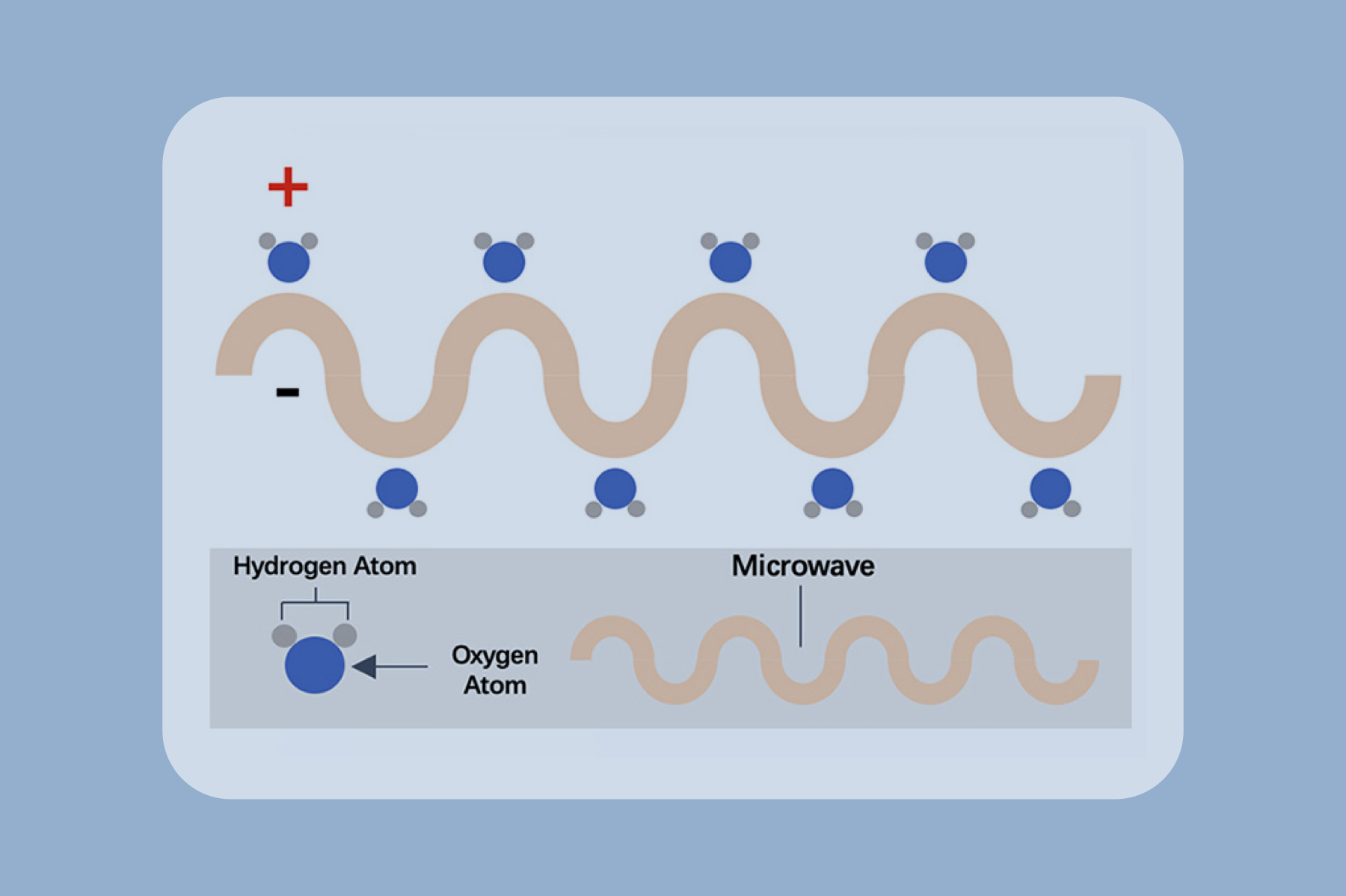
6. Simon, C. J., Dupuy, D. E., & Mayo-Smith, W. W. (2005). Microwave ablation: principles and applications. Radiographics, 25(suppl_1), S69-S83.

7. Liu, J. X., Li, J. Y., Zhao, X. Y., Zhang, Q. H., Cao, Y., Huang, X. J., ... & Yang, S. S. (2019). Transvaginal ultrasound-and laparoscopy-guided percutaneous microwave ablation for adenomyosis: preliminary results. International Journal of Hyperthermia, 36(1), 1232-1237.

8. Lin, M., He, J., Lyu, G., Li, Z., Li, X., Qiu, S., ... & Li, S. (2023). Combined transabdominal and transvaginal ultrasound-guided percutaneous microwave ablation of uterine myomas: an effective monitoring technique. International Journal of Hyperthermia, 40(1), 2154576.

9. Shunshi, Y., Li, J., Li, J., Huang, L., Chen, Y., Zhao, X., ... & Zhang, Q. (2023). Transvaginal ultrasound-and laparoscopy-guided percutaneous microwave ablation for adenomyosis has short-and long-term benefits: a single-center study. International Journal of Hyperthermia, 40(1), 2233713.

10. Zhang, J., Feng, L., Zhang, B., Ren, J., Li, Z., Hu, D., & Jiang, X. (2011). Ultrasound-guided percutaneous microwave ablation for symptomatic uterine fibroid treatment-a clinical study. International Journal of Hyperthermia, 27(5), 510-516.

11. Liu, H., Zhang, J., Han, Z. Y., Zhang, B. S., Zhang, W., Qi, C. S., ... & Xu, R. F. (2016). Effectiveness of ultrasound-guided percutaneous microwave ablation for symptomatic uterine fibroids: a multicentre study in China. International Journal of Hyperthermia, 32(8), 876-880.

12. Beermann, M., Jonsdottir, G., Cronisoe, A., Hasselrot, K., & Kopp Kallner, H. (2022). Long term follow-up of uterine fibroids treated with microwave ablation: an up to 3-year observational study of volume, regrowth, and symptoms. International Journal of Hyperthermia, 39(1), 1158-1163.

13. Xia, M., Jing, Z., Zhi-Yu, H., Jian-Ming, C., Hong-Yu, Z., Rui-Fang, x., ... & Bao-Wei, D. (2014). Feasibility study on energy prediction of microwave ablation upon uterine adenomyosis and leiomyomas by MRI. The British Journal of Radiology, 87(1040), 20130770.


  • Address: Building 3 Phase 2 Accelerator, No.11 Yaogu Avenue, Nanjing, Jiangsu, China
    Customer Service: service@canyonmedical.com.cn
  • Email: sales@canyonmedical.com.cn
    Postcode: 210032